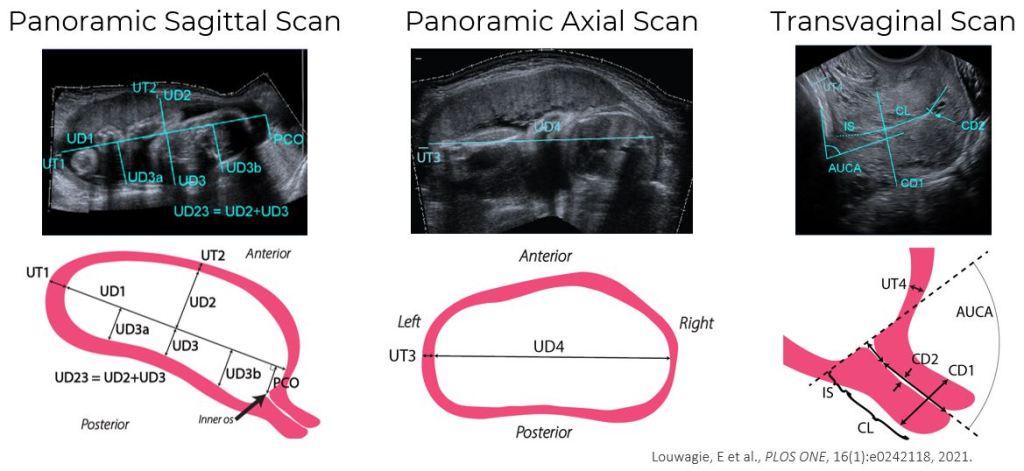
Ultrasonic dimension measurements of the maternal anatomy are collected, characterizing the overall size and shape of the uterus and cervix.
Maternal ultrasonic dimension measurements are entered into a design table in Solidworks, which automatically generates the parametric patient-specific geometry. Louwagie, E et al., PLOS ONE, 16(1):e0242118, 2021.
A typical simulation finite element result of an intrauterine pressure applied to the fetal membrane, showing the 1st principal Lagrange strain in the uterus and cervix. As the fetal membrane pushes into the cervical canal, the internal os is both compressed and begins to stretch open. The tissue at the uterocervical junction and lower uterine segment are also compressed and stretched. At the end of the simulation, the regions of greatest strain are the uterocervical junction and internal os.