In-Silico Models of In-Vivo Cervical Stiffness Measurements for Improving Preterm Birth Prediction
Adriana Delagarza, Erin Louwagie, Abigail Laughlin, Jacqueline Hairston, Mirella Mourad, Michael House, and Kristin Myers
Background
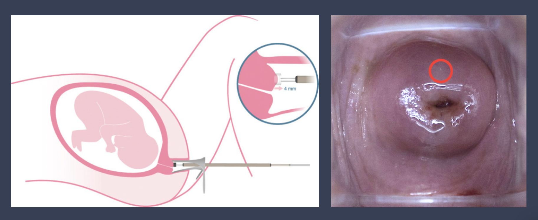
Cervical Aspirator (Pregnolia System)
Figure 1: (Left) Schematic of Pregnolia cervical aspiration device [1] and (Right) Visualization of probe placement on cervical tissue during speculum examination
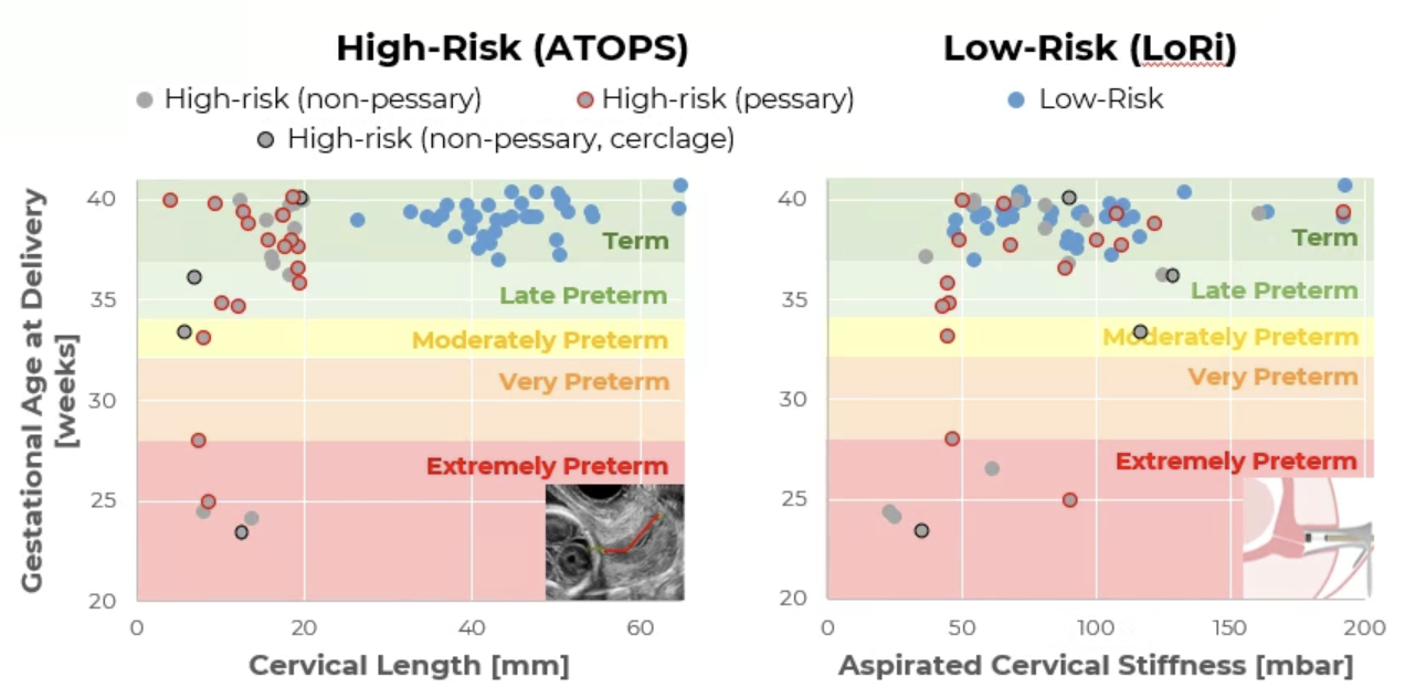
Figure 2: Comparison of correlations between (Left) cervical length and birthing outcome vs. (Right) aspirated cervical stiffness measurement using Pregnolia and birthing outcome
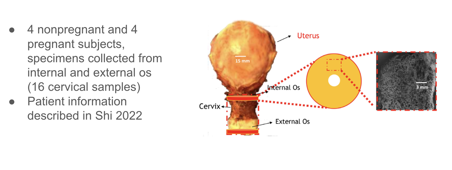
Figure 4: Data collection procedure for cervical model material properties [2]
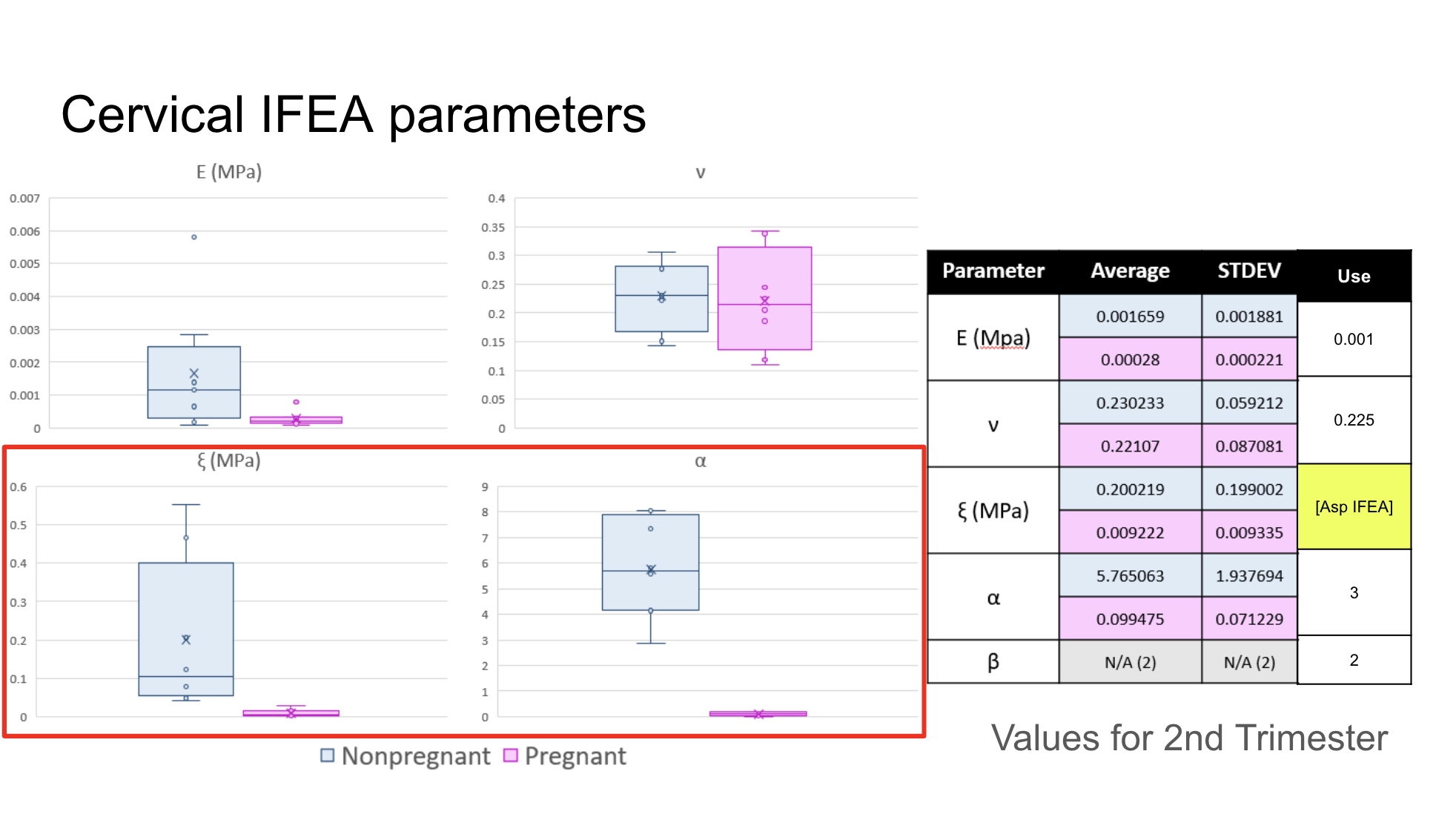
Figure 5: Mechanical parameters of cervical tissue for input into FEBio Studio. Cervical tissue was modeled as a composite fibrous material, with a neo-Hookean ground substance and embedded continuous spherically distributed fiber network, fit to existing tension and compression data. [2]
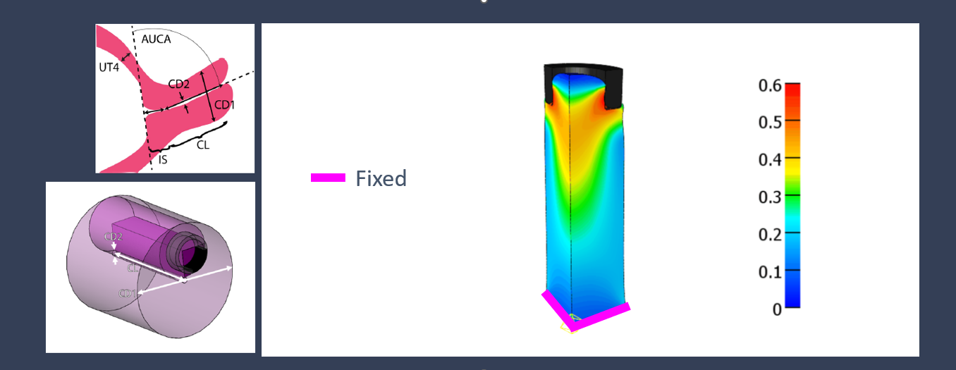
Model Setup
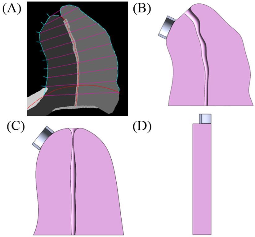
Figure 6: (A) Measurements taken from ultrasound images of the cervix, as well as cervical geometric fidelity modeled in SolidWorks for (B) high-, (C) mid-, and (D) low-fidelity simulations.
Figure 7: Head-on visualization of probe placement on the 12 o’clock position on the cervix.
Sensitivity Study: Boundary Conditions
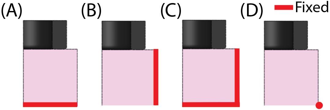
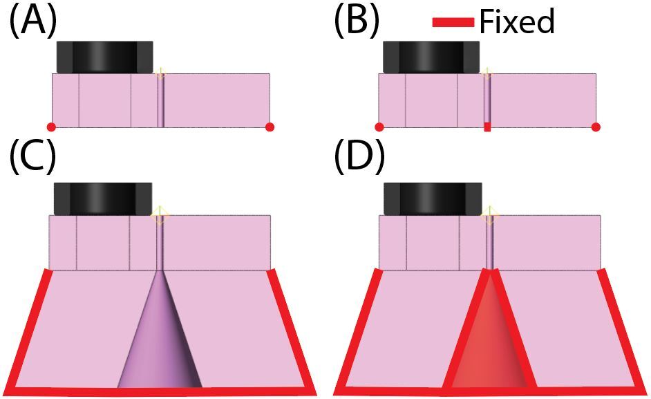
Figure 8: Outer faces of the cervix fixed at different points for investigation of boundary conditions on LF model
Sensitivity Study: Funnel Boundaries and Angle
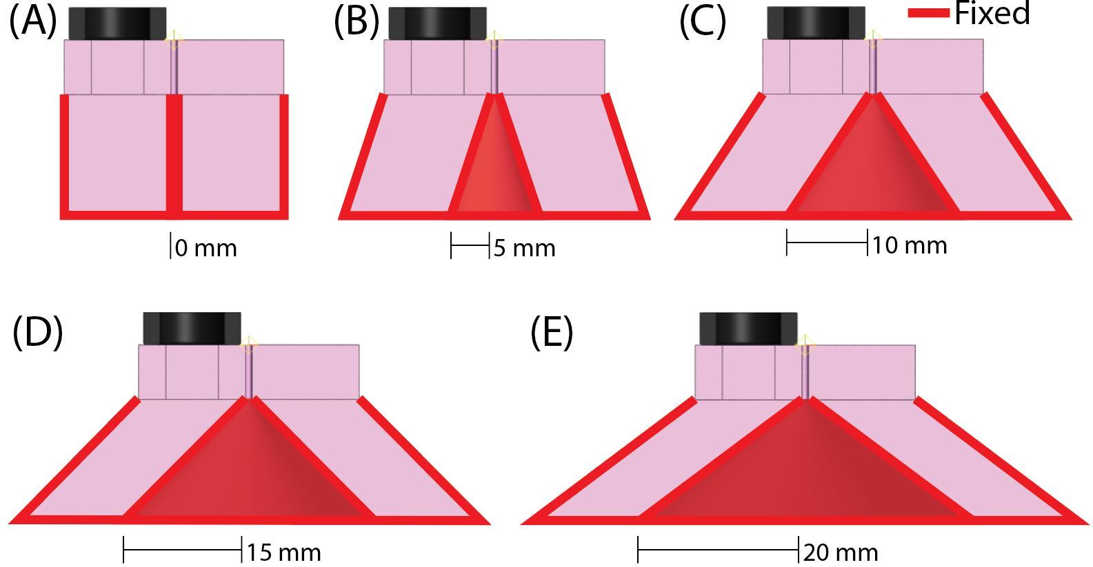
Figure 9: Sensitivity study of LF cervix with closed, fixed funnel and open, fixed funnel
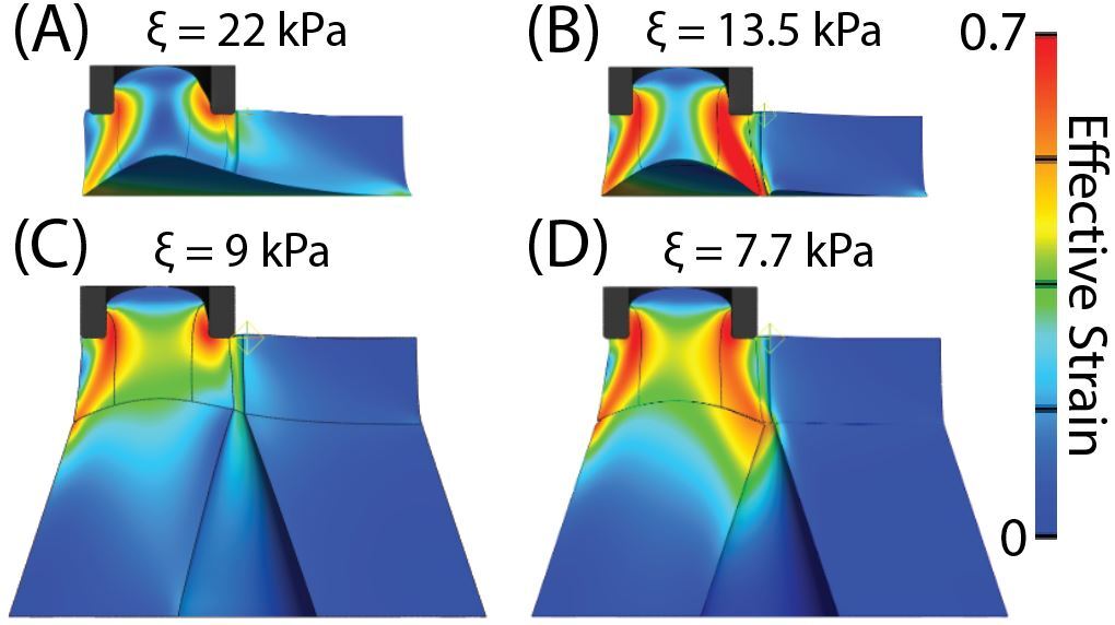
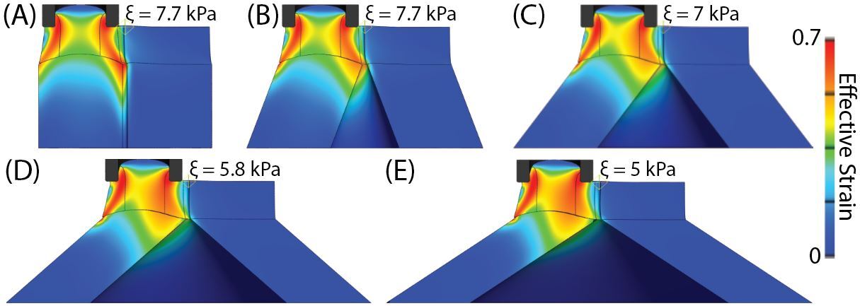
Figure 10: Cervical funnel changes and their effects on computed cervical stiffness values
Results
Figure 11: Effective Lagrange Strain during aspiration for LF model of low-risk patient
Figure 12: Effective Lagrange Strain during aspiration for MF model of low-risk patient
Figure 13: Effective Lagrange Strain during aspiration for HF model of low-risk patient
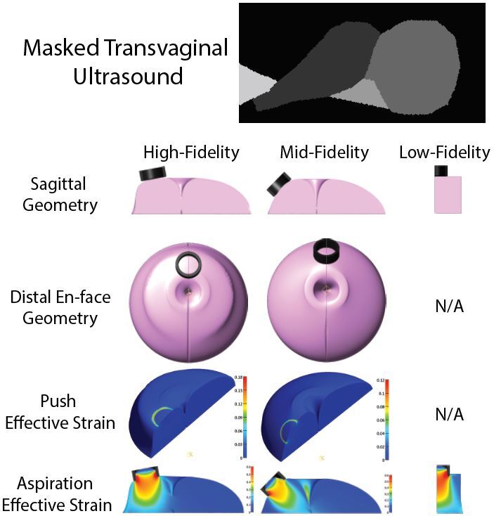
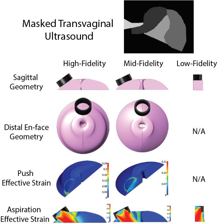
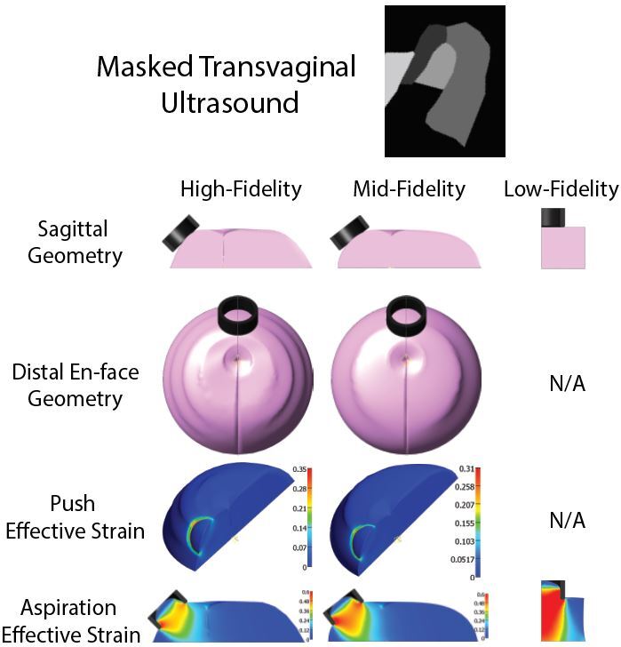
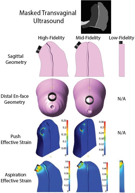
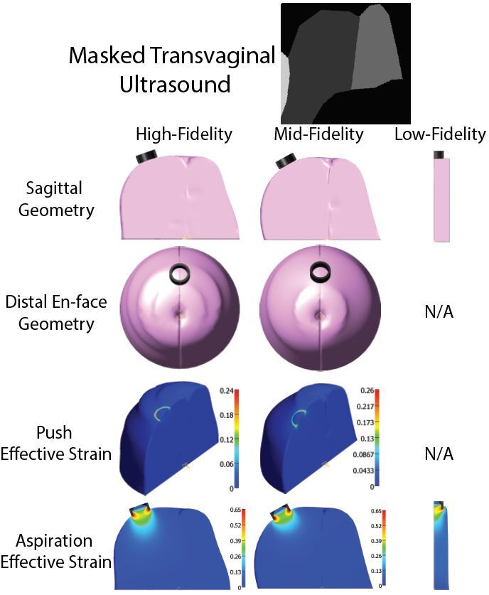
Figure 14: Masked ultrasound image, geometric fidelity changes, and strain heat maps for Patient 1 (High Risk)
Figure 15: Masked ultrasound image, geometric fidelity changes, and strain heat maps for Patient 2 (High Risk)
Figure 16: Masked ultrasound image, geometric fidelity changes, and strain heat maps for Patient 6 (High Risk)
Figure 17: Masked ultrasound image, geometric fidelity changes, and strain heat maps for Patient 7 (Low Risk)
Figure 18: Masked ultrasound image, geometric fidelity changes, and strain heat maps for Patient 16 (Low Risk)
Figure 19: Masked ultrasound image, geometric fidelity changes, and strain heat maps for Patient 32 (Low Risk)
Figure 20: Improved correlation between computed cervical stiffness with aspiration and birthing outcome
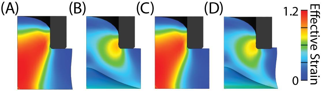
Figure 21: Strain heat maps for boundary condition study
Figure 22: Strain heat maps for cervical canal boundary condition study
Figure 23: Strain heat maps for changing cervical funnel angle study
References
[1] Pregnolia System – Cervical stiffness assessment: Instructions for Use, P/N 100041-H, Pregnolia AG, Schlieren, CH, 2024. Available: https://en.pregnolia.com/gebrauchsanweisung [2] Shi, L et al., “Three-dimensional anisotropic hyperelastic constitutive model describing the mechanical response of human and mouse cervix”, Acta Biomaterialia, 150(15):277-294, 2022.